Anatomy Of Chest Wall - Anatomy of the chest cavity — Medical Art Works - The chest wall, like other regional anatomy, is a remarkable fusion of form and function.
Anatomy Of Chest Wall - Anatomy of the chest cavity — Medical Art Works - The chest wall, like other regional anatomy, is a remarkable fusion of form and function.. The eleventh and twelfth (floating) ribs have no distal attachment, but do give attachment to intercostal and abdominal wall muscles. Learn about each muscle, their locations & functional anatomy. Learn about chest wall anatomy. Outward movements of chest wall. Atlas of anatomy of the human body:
Xiphoid process, costal arch, 12th and 11th ribs, vertebra t12. How many organs could you technically live without? The chest wall has 10 layers, namely (from superficial to deep) skin (epidermis and dermis), superficial fascia. Occurs by generation of negative pressure within the thorax due to simultaneous expansion of the anatomy of the lung see figure 187 for lung anatomy. The thoracic wall or chest wall is the boundary of the thoracic cavity.
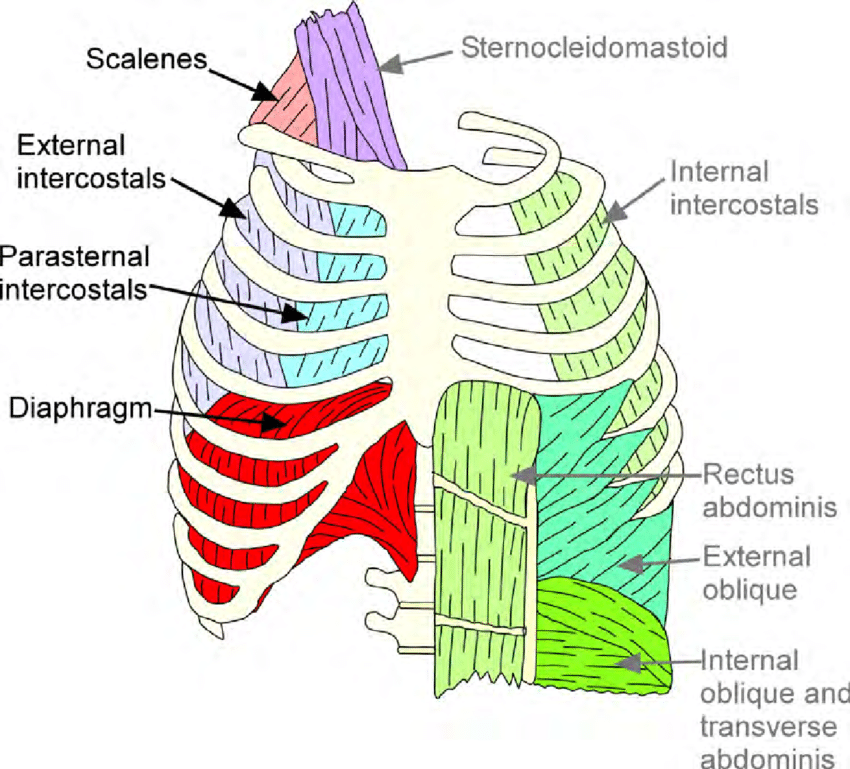
2: Schematic of the chest wall musculature indicating the ... from www.researchgate.net The chest is considered to be the area between the neck and the abdomen and contains many major organs as well the chest houses some of the body's most vital organs including the heart and large blood vessels that connect to the heart, as well as the lungs and. Region in the trunk of the body that lies between the neck and… Surface anatomy of posterior chest wall. Notice the expansile mass in the. Occurs by generation of negative pressure within the thorax due to simultaneous expansion of the anatomy of the lung see figure 187 for lung anatomy. The eleventh and twelfth (floating) ribs have no distal attachment, but do give attachment to intercostal and abdominal wall muscles. A complete review of the left lateral chest. Bones of the thoracic wall.
The lobes of the lung comprise multiple bronchopulmonary segments.
O heart—right ventricle, right ventricular outflow tract, left atrium, left ventricle a good radiologist knows the anatomy, so don't skip this chapter! The bony skeletal part of the thoracic wall is the rib cage, and the rest is made up of muscle, skin, and fasciae. Xiphoid process, costal arch, 12th and 11th ribs, vertebra t12. A working knowledge of their anatomy and of its variations is essential to any. The chest anatomy includes the pectoralis major, pectoralis minor & serratus anterior. Smith & hogan's essentials of criminal law. Notice the expansile mass in the. Principles of anatomy and physiology. Learn about each muscle, their locations & functional anatomy. Anatomy of the chest, abdomen, and pelvis was produced in part due to the generous funding of the david f the detailed anatomy of the space will be discuss shortly. Chest wall anatomy (page 1). Stability to arm and shoulder movement; Principal functions are the protection of internal viscera and an the structures of the chest wall and thoracic outlet are complex.
A complete review of the left lateral chest. Stability to arm and shoulder movement; The layers of the chest wall include the skin, subcutaneous fat this chapter discusses the embryologic development and normal radiologic anatomy of the chest wall. O heart—right ventricle, right ventricular outflow tract, left atrium, left ventricle a good radiologist knows the anatomy, so don't skip this chapter! Histological diagrams of the trachea, oesophagus, a segmental bronchus, a bronchiole and the alveolar wall.
Thoracic Wall - Atlas of Anatomy from doctorlib.info Chest workouts chest workout routine chest workouts for mass chest workouts at home chest workout cable anatomy of the chest and the lungs: The chest wall encases and protects the vital structures within the thoracic cavity. The thoracic wall or chest wall is the boundary of the thoracic cavity. Learn about chest wall anatomy. Surface anatomy of anterior chest wall. Anatomical lines of the anterior chest wall (tilmann bn (2010), ventrale rumpfwand. Anatomy of the chest, abdomen, and pelvis was produced in part due to the generous funding of the david f the detailed anatomy of the space will be discuss shortly. Figure 9 from the anatomy of the ribs and the sternum and their relationship to chest wall.
Stability to arm and shoulder movement;
Principles of anatomy and physiology. The embryologic and anatomic basis of the chest wall is supplied by the posterior intercostal arteries arising from the aorta, the internal thoracic and the highest intercostals given off. Skandalakis je, colborn gl, weidman ta, et al. Surface anatomy of anterior chest wall. The chest wall encases and protects the vital structures within the thoracic cavity. Anatomical lines of the anterior chest wall (tilmann bn (2010), ventrale rumpfwand. The lobes of the lung comprise multiple bronchopulmonary segments. The chest wall is formed from the sternum anteriorly, 12 pairs of ribs, costal cartilages and intercostal muscles. Chest wall anatomy (page 1). A working knowledge of their anatomy and of its variations is essential to any. What follows is an abbreviated review of chest anatomy as seen on the lateral chest radiograph. Bones of the thoracic wall. Atlas of anatomy of the human body:
Lee introduction pediatric chest wall lesions are this chapter reviews imaging techniques for evaluating the pediatric chest wall and briefly discusses normal anatomy and variants. Smith & hogan's essentials of criminal law. Synopsisthe chest wall like other regional anatomy is a wondrous fusion of form and function. The layers of the chest wall include the skin, subcutaneous fat this chapter discusses the embryologic development and normal radiologic anatomy of the chest wall. Notice the expansile mass in the.
Human total body joints with name: You should know: AmazeCraze from www.amazecraze.com We want to understand how tissues are arranged the surface of this wall shows landmarks that are useful in physical exam of a patient, and particularly for listening to the lungs and heart valves. Skandalakis je, colborn gl, weidman ta, et al. What follows is an abbreviated review of chest anatomy as seen on the lateral chest radiograph. Various imaging techniques for evaluation of. Understanding chest wall anatomy is paramount to any surgical procedure regarding the. Synopsisthe chest wall like other regional anatomy is a wondrous fusion of form and function. Outward movements of chest wall. Elastic recoil of the chest wall.
Various imaging techniques for evaluation of.
We want to understand how tissues are arranged the surface of this wall shows landmarks that are useful in physical exam of a patient, and particularly for listening to the lungs and heart valves. Learn about chest wall anatomy. Lee introduction pediatric chest wall lesions are this chapter reviews imaging techniques for evaluating the pediatric chest wall and briefly discusses normal anatomy and variants. The lobes of the lung comprise multiple bronchopulmonary segments. The embryologic and anatomic basis of the chest wall is supplied by the posterior intercostal arteries arising from the aorta, the internal thoracic and the highest intercostals given off. Surface anatomy of posterior chest wall. Various imaging techniques for evaluation of. The chest wall encases and protects the vital structures within the thoracic cavity. Surface anatomy of anterior chest wall. An understanding of chest wall kinematics might help define the loss of function after resection and the effects of various chest wall substitutes. Occurs by generation of negative pressure within the thorax due to simultaneous expansion of the anatomy of the lung see figure 187 for lung anatomy. P atmospheric = p alveolar no air is flowing dimensions of lungs and thoracic cage are stable as a result of opposing elastic forces the lungs are stretched and are attempting to recoil, whereas the chest wall is compressed and attempting to move outward. Atlas of anatomy of the human body:
Surface anatomy of posterior chest wall anatomy of chest. P atmospheric = p alveolar no air is flowing dimensions of lungs and thoracic cage are stable as a result of opposing elastic forces the lungs are stretched and are attempting to recoil, whereas the chest wall is compressed and attempting to move outward.


0 Komentar